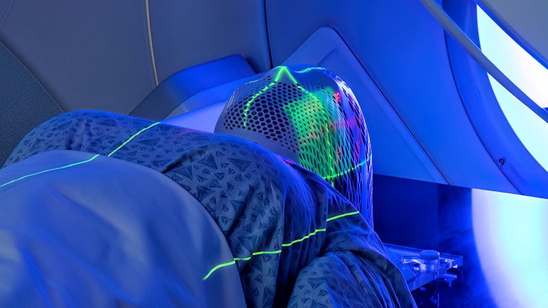
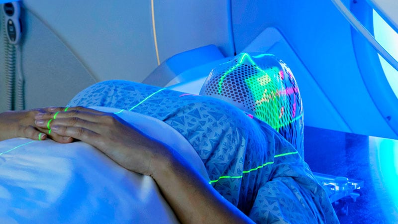
Post-treatment imaging at 6 months has better sensitivity and specificity for follow-up of head and neck (H&N) cancer patients compared with imaging at 3 months post-treatment, according to research published in the Journal of Cranio-Maxillofacial Surgery.
The retrospective study was performed for all H&N cancers treated with curative intent at the Royal Derby Hospital. Data collected included demographic information, site of primary cancer, staging, treatment provided, type of follow-up imaging performed, and results of follow-up imaging. Inclusion in the study was for oral, oropharyngeal, and hypopharyngeal cancers treated with curative intent, asymptomatic patients, those who had follow-up imaging within 6 months of treatment and those followed up for at least 2.5 years since treatment.
A total of 140 patients were included in the study. One in four patients had evidence of recurrent/metastatic disease on imaging, 60% of which were identified within six months of treatment. The majority (60%) of failures were due to distant metastases.
The sensitivity and specificity of both MRI and PET-CT was higher at 6 months post-treatment compared with 3 months post-treatment. Overall, the sensitivity and specificity of identifying treatment failure with PET-CT and MRI within 3-6 months post-treatment were 94.7% and 83.5% and 60% and 85.7%, respectively.
The authors concluded that, in patients with oral, oropharyngeal, and hypopharyngeal cancers with no clinical symptoms of recurrent disease, follow up PET CT is more sensitive than MRI in identifying treatment failure, both locoregional and distant.